
Modern dentistry has entered an era where precision, visibility and technology work together to improve outcomes. Root Canal Treatment is no longer limited to what the naked eye can see. With the use of advanced dental microscopes, this well known procedure has evolved into a more controlled, detailed and data driven intervention. In its simplest form, Root Canal Treatment under the microscope focuses on enhancing visualization of the inner tooth anatomy, allowing clinicians to explore areas that were once difficult to access or even impossible to notice.
From a patient perspective, Root Canal Treatment often carries uncertainty because it deals with the hidden structures of the tooth. Microscopic assistance changes this dynamic. It turns a traditionally tactile procedure into a visually guided one. This shift is not only technical but conceptual. Dentistry moves closer to microsurgery standards, where millimeter level accuracy matters and small details influence long term results.
ClinicHI follows this modern approach by integrating microscope assisted Root Canal Treatment into its clinical philosophy, emphasizing analysis, documentation and procedural clarity rather than assumptions.
How Root Canal Treatment Changes with Dental Microscopes
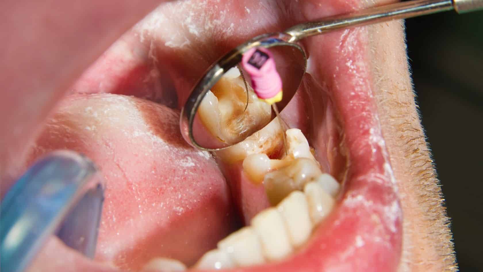
When comparing conventional techniques with microscopic Root Canal Treatment, the most striking difference is magnification. Dental microscopes can provide magnification levels ranging from 6x to over 25x. This allows the clinician to clearly identify canal entrances, micro cracks and anatomical variations. In traditional Root Canal Treatment, canal detection relies heavily on experience and tactile feedback. Under the microscope, visual confirmation becomes central. Subtle color changes in dentin, hidden accessory canals and calcified structures are more easily distinguished. This visual data supports decision making rather than replacing it. Another important transformation lies in illumination. Microscopes provide coaxial light that reaches deep into the canal system. This is especially relevant in Root Canal Treatment cases involving molars, where complex anatomy is common. Enhanced light reduces visual fatigue and increases procedural consistency across sessions.The Science Behind Microscopic Root Canal Treatment
From a scientific perspective, Root Canal Treatment under magnification aligns with principles of minimally invasive dentistry. Research in endodontics consistently highlights the relationship between visibility and procedural accuracy. Better visualization correlates with improved detection of untreated canals, which is a known factor in retreatment cases. Microscopic Root Canal Treatment also supports documentation and analysis. Procedures can be recorded in high resolution, allowing for post treatment review and academic evaluation. This creates a feedback loop where clinical outcomes inform future protocols. Another scientific advantage involves material interaction. Sealers, obturation materials and bonding agents behave differently depending on canal cleanliness and shape. By improving visual control during Root Canal Treatment, clinicians can better assess canal walls and material adaptation without relying solely on radiographic interpretation.Root Canal Treatment and Complex Tooth Anatomy
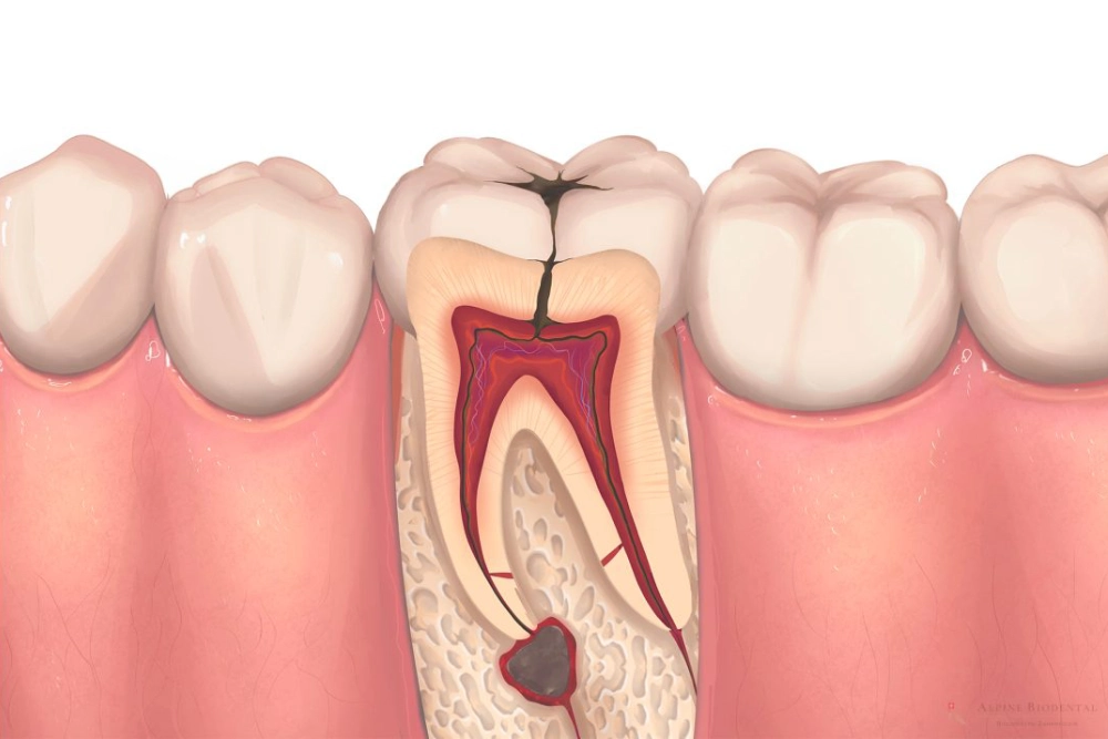
Human teeth rarely follow textbook anatomy. Variations are common, particularly in premolars and molars. Root Canal Treatment under the microscope reveals this complexity in real time rather than after complications arise. Accessory canals, isthmuses and apical deltas often remain undetected in standard procedures. Microscopic Root Canal Treatment increases the likelihood of identifying these structures during the initial intervention. This is not about guaranteeing outcomes but about reducing unknown variables. Understanding anatomy visually also supports conservative access cavity design. By preserving more tooth structure, microscopic Root Canal Treatment aligns with restorative principles that value long term tooth integrity over aggressive removal.Technology Integration in Root Canal Treatment Workflows
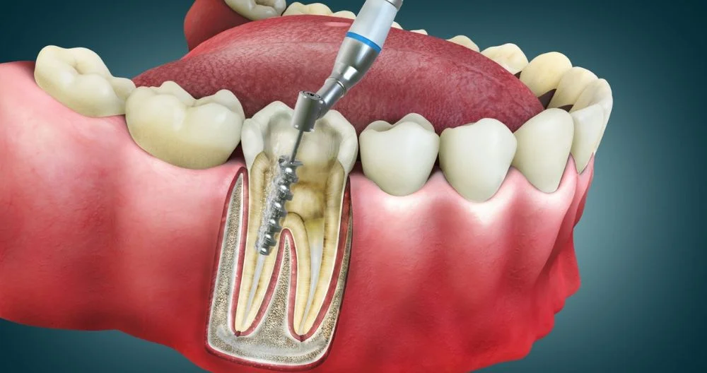
Microscope assisted Root Canal Treatment does not operate in isolation. It is usually part of a broader digital workflow that includes cone beam imaging, ultrasonic instruments and advanced irrigation systems. Each component adds a layer of information or control. For example, ultrasonics used under microscopic guidance allow precise dentin removal around calcified canals. In conventional Root Canal Treatment, this step may involve estimation rather than visualization. The combination of sound, vibration and magnification creates a multi sensory approach to canal negotiation. ClinicHI integrates these technologies to ensure that Root Canal Treatment is approached as a system rather than a single procedure. The microscope acts as a central reference point within this system.
Root Canal Treatment Under the Microscope and Clinical Decision Making
Decision making in Root Canal Treatment is influenced by what can be seen, measured and documented. Microscopic assistance provides real time visual feedback that informs each step, from access cavity preparation to obturation. This does not eliminate uncertainty but reframes it. Instead of relying on indirect signs, clinicians can observe canal morphology directly. This is particularly relevant in retreatment scenarios, where previous materials or procedural changes alter the internal landscape of the tooth. Microscopic Root Canal Treatment also supports ethical transparency. Visual records can be used to explain procedural findings, reinforcing trust without making definitive promises or health claims.Materials, Precision and Root Canal Treatment Outcomes
Materials used in Root Canal Treatment have evolved significantly, but their effectiveness depends on how accurately they are applied. Microscopic visualization improves precision during irrigation, shaping and filling phases. For instance, debris removal from canal walls is easier to assess under magnification. This does not imply perfection but enhances awareness. Similarly, obturation techniques benefit from visual confirmation of material flow and adaptation. In scientific literature, Root Canal Treatment success is often discussed in probabilistic terms rather than absolutes. Microscopic assistance contributes to this discussion by reducing technical variability, which is one of the known factors affecting procedural consistency.Why Microscopic Root Canal Treatment Reflects Modern Dentistry
Modern dentistry emphasizes evidence, documentation and continuous improvement. Root Canal Treatment under the microscope reflects these values by transforming an experience based procedure into an observable and analyzable one. It also aligns with patient centered communication. When procedures are visualized and recorded, explanations become clearer and less abstract. This supports informed understanding without offering medical advice or definitive outcomes. ClinicHI positions microscopic Root Canal Treatment as part of its broader commitment to technological literacy and clinical transparency rather than as a standalone feature.Root Canal Treatment as a Field of Ongoing Research
Endodontics continues to evolve, and Root Canal Treatment under magnification is an active area of research. Studies explore topics such as canal detection rates, procedural ergonomics and long term tooth preservation. The microscope serves as both a clinical and research tool. It bridges the gap between academic findings and everyday practice. By observing details that were previously inferred, microscopic Root Canal Treatment contributes to a more nuanced understanding of tooth biology. This research driven approach reinforces the idea that Root Canal Treatment is not static. It adapts as tools, materials and analytical methods advance.Final Thoughts on Root Canal Treatment Under the Microscope
Microscope assisted Root Canal Treatment represents a shift in how dental professionals interact with the unseen structures of the tooth. It emphasizes visibility, analysis and documentation over assumption. Rather than redefining the procedure itself, it refines how each step is performed and evaluated. This refinement supports scientific inquiry, clinical consistency and transparent communication. At ClinicHI, Root Canal Treatment under the microscope is viewed as part of a broader evolution toward precision based dentistry, where knowledge grows through observation and continuous learning rather than definitive claims.Get Your Free Consultation
Have questions? Leave your details and we'll get back to you as soon as possible.
Get Free Consultation